Home » Without Label » Cross Section Of A Bone : Krames Online - Crecimiento del esqueleto pediátrico : After a fracture, woven bone forms initially and is gradually replaced by lamellar bone during a process known as bony substitution.
Cross Section Of A Bone : Krames Online - Crecimiento del esqueleto pediátrico : After a fracture, woven bone forms initially and is gradually replaced by lamellar bone during a process known as bony substitution.
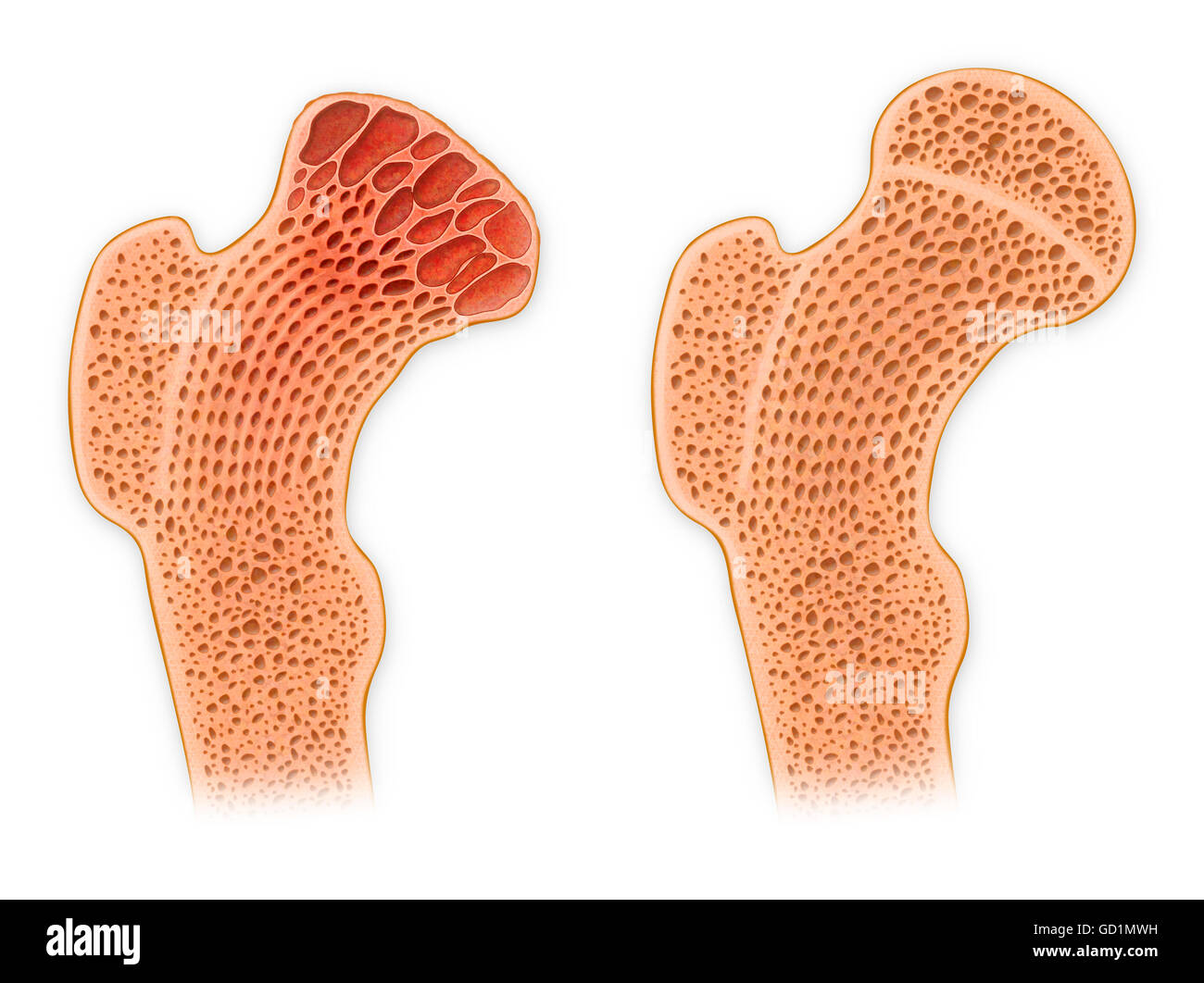
Cross Section Of A Bone : Krames Online - Crecimiento del esqueleto pediátrico : After a fracture, woven bone forms initially and is gradually replaced by lamellar bone during a process known as bony substitution.. Smooth muscle and endothelium in a muscular artery wall, (magnification x100). Marrow in the shaft of long bones is typically yellow, with red marrow in the head through the cancellous bone. While it is not as hard as compact bone, spongy bone plays an important role of protecting the marrow where blood cells are produced. Each bone in your body is made up of three main types of bone material: Foot bone anatomy x ray 12 photos of the foot bone anatomy x ray foot bone anatomy x ray, bone, foot bone anatomy x ray.
Cross section of mandible at first molar region showing cortical and spongy bone basic concepts in osteogenesis. Smooth muscle and endothelium in a muscular artery wall, (magnification x100). The geometrical properties generated from the ct image included as follows: In addition, cortical bone thickness at anterior, posterior, medial, and lateral parts of the bone section was measured. At the outer regions of the section, you can see a dense, thick layer of compact bone.
Bone Cross Section / Bone Cross Section High Res Stock ... from c8.alamy.com The inner portion of the bone is composed of trabecular bone and the intervening bone marrow. Related posts of cross section of a long bone muscles and bones in the arm. Marrow in the shaft of long bones is typically yellow, with red marrow in the head through the cancellous bone. Cross section of mandible at first molar region showing cortical and spongy bone basic concepts in osteogenesis. The upper (biting) surfaces of the tooth are at top, with the lower sections (bottom) embedded in the gums and jaw bone (not shown). And why does the marrow stop where it does, and so sharply? The outlined area is a cross section of an osteon of compact bone. In three dimensions an osteon is cylindrical in shape.
Assuming the cross section of the bone at c to be annular and knowing that its outer diameter is 25 mm, determine the inner diameter of the bone's cross section at c.
The large dark spots are passages for blood vessels and nerves. Related posts of cross section of human bone diagram foot bone anatomy x ray. In addition, cortical bone thickness at anterior, posterior, medial, and lateral parts of the bone section was measured. It consists of two layers; At the outer regions of the section, you can see a dense, thick layer of compact bone. Marrow in the shaft of long bones is typically yellow, with red marrow in the head through the cancellous bone. Body size standardization was done, using the following equations: Concentric layers of bone cells (osteocytes) and bone matrix surround the central canal. Foot bone anatomy x ray 12 photos of the foot bone anatomy x ray foot bone anatomy x ray, bone, foot bone anatomy x ray. As the names suggest compact bone looks compact and the spongy bone looks like sponges. They also produce various blood cells, store minerals, and provide support for mobility in femur head showing trabecular bone : The surface features of bones vary considerably, depending on the function and location in the body. Each bone in your body is made up of three main types of bone material:
Bone markings the surface features of bones vary considerably, depending on the function and location in the body. The large dark spots are passages for blood vessels and nerves. They also produce various blood cells, store minerals, and provide support for mobility in femur head showing trabecular bone : There are three general classes of bone. In the center of each osteon is the central canal, a space that houses blood vessels and nerves that supply bone.
Anatomy Tissue Practical Review Flashcards | Easy Notecards from www.umich.edu Body size standardization was done, using the following equations: Bone matrix and cells bone matrix osseous tissue is a connective tissue and like all connective tissues contains relatively few cells and large amounts of extracellular matrix. Foot bone anatomy x ray 12 photos of the foot bone anatomy x ray foot bone anatomy x ray, bone, foot bone anatomy x ray. Related posts of bone cross section labeled. The central tubular region of the bone, called the diaphysis, flares outward near the end to form the metaphysis, which contains a largely cancellous, or spongy, interior. In addition, cortical bone thickness at anterior, posterior, medial, and lateral parts of the bone section was measured. Table 1 describes the bone markings, which are illustrated in (figure 4). Compact bone is the outer layer and the spongy bone forms the inner layer.
The geometrical properties generated from the ct image included as follows:
The geometrical properties generated from the ct image included as follows: An outer 'fibrous layer' containing mainly fibroblasts, and an inner 'cambium layer' containing progenitor cells. Foot bone anatomy x ray 12 photos of the foot bone anatomy x ray foot bone anatomy x ray, bone, foot bone anatomy x ray. In addition, cortical bone thickness at anterior, posterior, medial, and lateral parts of the bone section was measured. Related posts of bone cross section labeled. Two types of bone tissues in cross section of a long bone : And why does the marrow stop where it does, and so sharply? Compact bone is the outer layer and the spongy bone forms the inner layer. There are trabeculae in spongy bone which gives its sponge like appearance. Related posts of cross section of human bone diagram foot bone anatomy x ray. The upper (biting) surfaces of the tooth are at top, with the lower sections (bottom) embedded in the gums and jaw bone (not shown). Related posts of cross section of a long bone muscles and bones in the arm. Would it be a good thing to show the epiphyseal plate?
The cell line involved in osteogenesis consists of preosteoblasts, osteoblasts, osteocytes and bone. Browse 4,294 bone cross section stock photos and images available, or search for human bone cross section to find more great stock photos and pictures. The compact bone is made up of osteon. Internal structure of a human long bone. And why does the marrow stop where it does, and so sharply?
Cartilage and Bone - Slide #13 from education.med.nyu.edu The upper (biting) surfaces of the tooth are at top, with the lower sections (bottom) embedded in the gums and jaw bone (not shown). The geometrical properties generated from the ct image included as follows: At the outer regions of the section, you can see a dense, thick layer of compact bone. It consists of two layers; Related posts of cross section of human bone diagram foot bone anatomy x ray. In a cross section of a bone, you can usually see two types of bone tissues. An outer 'fibrous layer' containing mainly fibroblasts, and an inner 'cambium layer' containing progenitor cells. Internal structure of a human long bone, with a magnified cross section of the interior.
Foot bone anatomy x ray 12 photos of the foot bone anatomy x ray foot bone anatomy x ray, bone, foot bone anatomy x ray.
Concentric layers of bone cells (osteocytes) and bone matrix surround the central canal. Would it be a good thing to show the epiphyseal plate? Related posts of cross section of a long bone muscles and bones in the arm. Related posts of cross section of human bone diagram foot bone anatomy x ray. After a fracture, woven bone forms initially and is gradually replaced by lamellar bone during a process known as bony substitution. Internal structure of a human long bone. Body size standardization was done, using the following equations: Bone is a dynamic biological tissue, composed of various metabolically active cells that are integrated into a rigid framework. Cross section of mandible at first molar region showing cortical and spongy bone basic concepts in osteogenesis. Compact bone is the outer layer and the spongy bone forms the inner layer. Now that you know what bones do, let's take a look at what they're made of and their anatomy. Browse 4,294 bone cross section stock photos and images available, or search for human bone cross section to find more great stock photos and pictures. In a cross section of a bone, you can usually see two types of bone tissues.